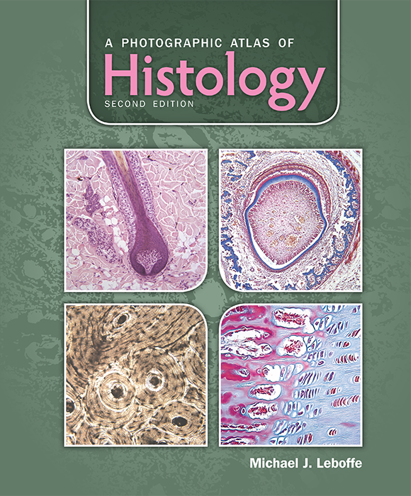
A Photographic Atlas of Histology, 2e
Michael J. Leboffe
This full-color photographic atlas is designed as a visual reference to accompany an undergraduate histology or human anatomy course. Hundreds of labeled photomicrographs give students a detailed look at the tissues of the human body.

Top Hat Interactive eText
requires a join code from instructor
$52
Interactive eText
does not require a join code
$42.00

Table of Contents for A Photographic Atlas of Histology, 2e
- Front Matter
- Ch 01: Introduction
- Ch 02: The Cell
- Ch 03: Epithelial Tissues
- Ch 04: Fibrous Connective Tissue
- Ch 05: Cartilage and Bone
- Ch 06: Blood and Bone Marrow
- Ch 07: Muscle Tissue
- Ch 08: Nervous Tissue and Organs of the Nervous System
- Ch 09: Special Sensory Organs
- Ch10: Endocrine System
- Ch 11: Integumentary System
- Ch 12: Cardiovascular System
- Ch 13: Lymphatic System